ICU resources for COVID-19, vitamin D and the immune system

Marik Protocol; Front Line COVID-19 Critical Care Working Group Protocol; Farid Jalali's beautiful detective work hypothesising about exactly what is happening in the lungs of COVID-19 patients; Vitamin D to stabilize the endothelium . . .

Robin Whittle rw@firstpr.com.au .   2020-04-09  Last major update 2023-06-29.
Updated 2021-11-12 regarding the first article below, Chauss et al. 2021, which is the peer-reviewed successor to the preprint McGregor et al. 2020. 
Updated 2023-06-29: "Autocrine" corrected to "intracrine" and a few updates and improvements to the Chauss et al. / McGregor et al. section.

The updates/ page lists all the significant updates to these cv19 pages.

To the main COVID-19 page of this site: http://aminotheory.com/cv19/ , concerning nutritional supplements - especially vitamin D and boron - for all adults and some children ASAP, to reduce or eliminate the tendency for their immune system to be weak and/or dysregulated (overly-aggressive, pro-inflammatory and so self-destructive) - which is what causes severe symptoms with COVID-19.   There you will find multiple articles recommending that everyone should have at least 40ng/ml (100nmol/l) 25OHD levels - and this starts with expectant mothers and their newborns.

If everyone in the world was already replete in vitamin D alone (with the current very poor boron status) then I believe that far fewer people would be dying or seriously harmed due to COVID-19.    In the absence of proper vitamin D levels, it is possible that boron repletion (such as 6mg a day, instead of the usual 1mg or so) would be helpful too.   See the main page for more on boron and immunity.

Obesity ../obesity/ would still be a problem, as would some particular patterns of genes ../#haplotypes - but the outcomes would be far less extreme than they are now, with many or most of these people seriously deficient in vitamin D and probably other nutrients.  Please see the ../obesity/  page for links to research concerning excess adipocytes expressing ACE2, releasing pro-inflammatory cytokines and existing as ectopic adipocytes in the cells of the alveoli.


Be sure to read the Disclaimer!


#2020-McGregor and #2021-Chauss

COVID-19 patients need sufficient 25-hydroxyvitamin D to ensure Th1 lymphocytes in their lungs shut down and so reduce inflammation

2023-06-29 note: 

While Chauss et al. refer to autocrine signaling, they are really referring to and researching intracrine signaling, since the receptor is in the cytosol of the cell in which its ligand - 1,25-dihydroxyvitamin D - is produced.  If the receptor was bound to the outside of the plasma membrane of the cell in which the ligand was produced, with these molecules leaving the cell and binding to those external receptors on the same cell, this would be autocrine signaling.

This mistake is easy to make - I thought it was autocrine signaling too.

See: https://vitamindstopscovid.info/02-intracrine/ .


I regard this as the most important article of all regarding the mechanisms of severe COVID-19 - and more generally of how most people's low 25-hydroxyvitamin D levels lead to weakened immune responses and so more severe symptoms from COVID-19 and numerous other infectious diseases.  For research on why the immune system needs at least 50 ng/mL 125 nmol/L circulating 25-hydroxyvitamin D to function properly, please see: https://vitamindstopscovid.info/00-evi/.

Autocrine vitamin D signaling switches off pro-inflammatory programs of Th1 cells
Daniel Chauss, Tilo Freiwald, Reuben McGregor, Bingyu Yan, Luopin Wang, Estefania Nova-Lamperti, Dhaneshwar Kumar, Zonghao Zhang, Heather Teague, Erin E. West, Kevin M. Vannella1, Marcos J. Ramos-Benitez, Jack Bibby, Audrey Kelly1, Amna Malik1, Alexandra F. Freeman, Daniella M. Schwartz, Didier Portilla1, Daniel S. Chertow, Susan John, Paul Lavender, Claudia Kemper, Giovanna Lombardi, Nehal N. Mehta, Nichola Cooper1, Michail S. Lionakis, Arian Laurence, Majid Kazemian and Behdad Afzali
Nature Immunology 2021-11-11
https://www.nature.com/articles/s41590-021-01080-3

This is the peer-review journal result of a preprint from July 2020:

An autocrine Vitamin D-driven Th1 shutdown program can be exploited for COVID-19
Reuben McGregor, (22 others), Majid Kazemian and Behdad Afzali. 2020-07-19  (Preprint being edited for publication.)
https://www.biorxiv.org/content/10.1101/2020.07.18.210161v1


#2021-Minton

As befits an article of this significance, there is a Research Highlight in Nature Reviews Immunology:

Vitamin D shuts down T cell-mediated inflammation
Kirsty Minton
Nature Reviews Immunology 2021-11-19
https://www.nature.com/articles/s41577-021-00663-3

Here are some comments on this Research Highlight

I am being pernickety since so much (general health and suppressing COVID-19 pandemic transmission, harm and death) depends on a clear understanding of vitamin D based autocrine intracrine signaling.  Chauss et al. is a tremendous addition to our understanding, but the whole vitamin D field is bedeviled by confused and confusing terminology, and to some degree by the mistaken belief that immune cells need more hormonal (blood-borne) 1,25-hydroxyvitamin D, when what they need is good supplies of 25-hydroxyvitamin D.
 (https://vitamindstopscovid.info/02-intracrine/#02-nothorm .)
Once the vitamin D based intracrine signaling system is activated by a sufficient level of extracellular complement factor C3b causing the 1-hydroxylase enzyme and VDR to be produced in the cytosol, this enzyme hydroxylates 25-hydroxyvitamin D (which must be present in the cytosol in sufficient quantities) to 1,25-dihydroxyvitamin D, each molecule of which promptly binds to a VDR molecule.  These bound complexes enter the nucleus where they bind to retinoid X receptors and the triple complexes initiate a complex series of histone, DNA and vitamin D response element interactions which alter how a large number of genes are transcribed to mRNA.  The transcription of dozens or hundreds of genes are up-regulated and dozens or hundreds are down-regulated.

In general principle these mechanisms are the same in all kinds of cell which utilise vitamin D intracrine signaling as part of how so each cell responds to its changing circumstances.  The particular details of what initiates the intracrine signaling system, and the specific details of which genes have their transcription up- and down-regulated, vary from one cell type to the next.  In the case of these Th1 lymphocytes, the researchers identified that successful completion of the intracrine signaling cascade resulted in the upregulation of 262 genes and the downregulation of 128.   Among the cellular changes which result: reduced production of pro-inflammatory IFNγ; increased production of anti-inflammatory IL-10 and IL6 and  increased production of transcription factors including JUN, BACH2 and STAT3.


#2020-McGregor-summary
The following section is based on McGregor et al. preprint.  I am not sure when I will have time to fully read and understand Chauss et al.  My initial impression is that it elaborates on but does not contradict what was in McGregor et al.

So please use the following summary of McGregor et al. as a rough guide to Chauss et al. subject to the following caveats:
  1. This summary is of a preprint, while the full article is more elaborate, refined and considered - with further experimental results.

  2. This summary was written by an electronic technician and computer programmer who knows only a tiny fraction of the immunology and cell biology which would be required to fully understand and appraise these articles.

The following box repeats my summary from the list of recent research ../#lr and in the 02-intracrine page (which originally referred to, erroneously, autocrine signaling - below the text is as updated on 2023-06-29).  Below the box is a more detailed summary of the cellular mechanisms described in this article.

Th1 lymphocytes isolated from the lungs of patients with severe COVID-19 symptoms have an intracrine (https://vitamindstopscovid.info/02-intracrine/) signaling pathway which should be activated by high levels of complement (WP), to turn these cells off their initial hyper-inflammatory program which produces pro-inflammatory IFNγ (interferon_gamma WP which has antiviral and anti-bacterial activity as well as stimulating inflammation: cell destruction such as by natural killer cells WP) and instead cause them to produce the anti-inflammatory cytokine IL-10.  (The cells always produce both these cytokines, but this transition to a shutdown, anti-inflammatory program, involves less IFNγ and a lot
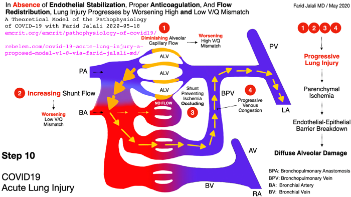
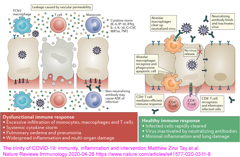
 more IL-10.)

However, this anti-inflammatory pathway is not working in the Th1 cells from patients with severe COVID-19, due solely to insufficient 25-hydroxyvitamin D = 25(OH)D = calcifediol for each cell's intracrine signaling system to function.  (Until 2021-03-01 I mistakenly stated that the Th1 cells initially produced IL-17 - and that the experimenters restored the Th1's anti-inflammatory pathway by adding 25(OH)D in-vitro.)

(The final Chauss et al. article stated that the cause was largely or entirely due to inadequate 25-hydroxyvitamin D)

This is a molecular and cellular explanation for why people with low 25-hydroxyvitamin D have self-destructive, wildly dysregulated, indiscriminate cell destroying, overly-inflammatory immune responses.  Such responses drive sepsis, severe influenza, Kawasaki disease (KD WP), Multisystem Inflammatory Syndrome (MIS discussion) and of course severe COVID-19.  (See Paul Marik's explanation https://www.evms.edu/covid-19/covid_care_for_clinicians/ of how it is the immune response, not the virus, which causes the escalation to severe symptoms and death.  See ../#2015-Stagi for research which shows KD children have very low 25(OH)D vitamin D levels.)

In severe COVID-19, severe inflammation in the lungs damages endothelial cells (the inner lining of blood vessels and capillaries WP) leading to hypercoagulative blood, causing microembolisms and larger clots all over the body, which cause most of hypoxia, lasting harm and death.

It is not known whether the cause of all the hyper-inflammatory immune system dysregulation - which causes some COVID-19 sufferers people to develop severe symptoms - is primarily the failure of these Th1 lymphocytes to switch from being pro- to anti-inflammatory, or whether this endothelial cell destruction etc. is also driven to a significant degree by similar failures in the intracrine signaling systems of many other types of regulatory and/or directly anti-pathogen immune cell and/or by direct actions of the virus.  However, the determination of the exact mechanism of failure in Th1 cells, in the context of such failures likely also occurring in other cell types, is an extraordinarily valuable contribution which deserves to be very widely known.

Low 
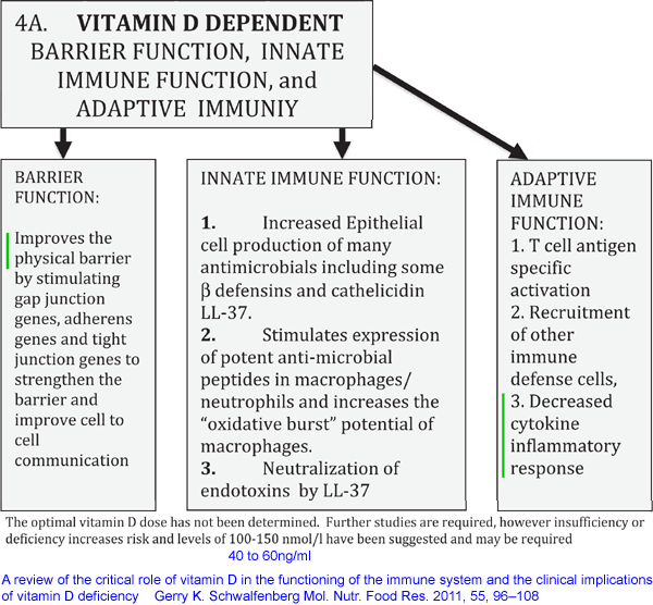
circulating 25-hydroxyvitamin D (produced in the liver from UV-B-generated and/or ingested vitamin D3 cholecalciferol) levels are well known to reduce the effectiveness of numerous direct, anti-pathogen, responses by the innate immune system cells and to hinder the creation of antibodies for adaptive immune responses.  These immune functions of 25-hydroxyvitamin D are due to it being needed, in the circulation, at higher levels than are sufficient for bone health (sufficient for the kidneys to produce their much lower concentration of circulating - and so hormonal - 1,25-dihyroxyvitamin D), to supply the intracrine / paracrine (inside the cell / to nearby cells) signaling systems of many or perhaps all types of immune cells.  All types of immune cell can express the vitamin D receptor - and this is for intracrine/paracrine signaling - not for responding to the much lower levels of circulating 1,25-dihydroxyvitamin D which regulates calcium-bone metabolism. https://vitamindstopscovid.info/02-intracrine/#02-nothorm .

See http://aminotheory.com/cv19/#2020-Fabbri [B] for why 40ng/ml or more 25OHD is required for these autocrine signaling systems to function properly.  See also the Quraishi et al. graph https://vitamindstopscovid.info/02-intracrine/#04-quraishi which suggests that innate immune cell responses which fight bacterial and perhaps fungal infections keep improving, presumably due to faster and stronger intracrine/paracrine signaling, as 25-hydroxyvitaminD levels rise, up to about 55ng/ml.

Please also see ../#25plusD3 for my suggestion of oral calcifediol (25-hydroxyvitamin D) plus vitamin D3 as the best treatment for hospitalised COVID-19 patients, since this raises circulating 25-hydroxyvitamin D to the levels needed for intracrine / paracrine signaling in a few hours, rather than in the several days to a week with, for instance, a 10 mg 400,000 IU bolus dose of vitamin D3.  See this 0.014 mg calcifediol per kg body weight recommendation in Prof. Wimalawansa's article in Nature, in 2022: https://www.mdpi.com/2072-6643/14/14/2997.

Very strong clinical evidence of the importance of rapidly raising circulating 25OHD levels hospitalised COVID-19 patients can be found the Cordoba calcifediol (25OHD) RCT: Castillo et al. 2020: ../#2020-Castillo .


Here is a more detailed summary of the cellular mechanisms reported in this article - but please remember that this is the best effort of an electronic technician and computer programmer, trying to summarize a dense cell biology preprint reporting on an extensive project conducted by 25 researchers.  What I have written below goes beyond a summary and includes elements of critique (regarding terminology) and some commentary - so please parse it carefully and refer to the article itself, rather than to what I write here, for your final decisions on the veracity and meaning of this research.

!! The graphic below refers to "autocrine" but it should be "intracrine" signaling.

McGregor et al. An autocrine Vitamin D-driven Th1 shutdown program can be exploited for COVID-19

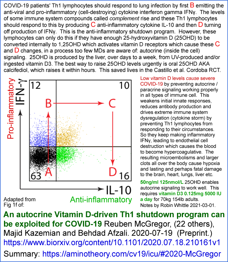
The wide positioning of the A, B, C and D on  the above graph exaggerates the relative changes in the degree to which these two cytokines are created. (The horizontal and vertical dimensions of Fig 1f are for the rates at which the genes for these two cytokines are transcribed, which is approximately proportional to the rates at which the cytokines themselves are produced.)

I wanted to know the relative concentrations of the two cytokines produced by the cells between the B activated, inflammatory, state and the D shutdown, anti-inflammatory state.  This can be estimated from each of two sets of data. 

Firstly, the graphs in Fig 2C show the levels of the two cytokines produced when there is zero 1,25-dihydroxyvitamin D (hereafter 1,25(OH)2D) supplied to the cells, and then the levels when three different concentrations of 1,25(OH)2D are supplied.  I judge the 10nM supply to be sufficient to fully activate the VDRs as they would be by normal, full, activation of the autocrine signaling system with sufficient 25-hydroxyvitamin D (hereafter 25(OH)D) supplied.  So, for instance, for the top IFNγ curve of Fig 2c, I can divide the ~5900 pg/ml shutdown D level (4th red dot from the left) by the ~9,800 pg/ml B initial, inflammatory, level and get a   D/B shutdown/inflammatory ratio of 0.6.  

Secondly I can do the same for Fig S5d (in the separate Supplementary Materials PDF) which represents the same things, but this time in response to the experimenters supplying 25(OH)D so the real, already activated, intracrine signaling system can operate, producing its own 1,25(OH)2D to activate the cell's VDRs.  Here, I chose the 3rd dot from the right, because I assumed, perhaps wrongly, that the 50nM (20ng/ml) 25(OH)D concentration was sufficient for the autocrine signaling system to operate fully.  For IFNγ in this graph I estimate 7100 / 9100 (why not 9800 as before adding 1,25(OH)2D?) = 0.78.

I estimate that the  D/B shutdown/inflammatory ratios are, from the two techniques respectively (first added 1,25(OH)2D and second added 25(OH)D) to be IFNγ 0.6 & 0.78, and IL-10 2.5 & 1.4 (ignoring the vertical scale discrepancy, which I assume is a typo).  Neither of these experimental techniques tell us exactly what quantities of the two cytokines Th1 cells produce in-vivo, but it is the best we can do. 

The normal, healthy, behaviour of Th1 lymphocytes to SARS-CoV-2 infection is as follows.  (Those isolated from the lungs of severe COVID-19 sufferers did not transition to the anti-inflammatory program, and kept producing the pro-inflammatory IFNγ, presumably due to lack of sufficient 25(OH)D in those patient's bloodstream and therefore interstitial fluid, or whatever fluids the cells were bathed in.)

This discussion concerns T helper lymphocytes (WP) which developed from Th0 program cells into Th1 types, were attracted to a site of infection - in this case the lungs of hospitalised COVID-19 patients - and there multiplied (AKA, I think, "differentiated") so they are present in much higher than numbers at this site than when they first arrived. 

These Th1 cells have been activated (described below, perhaps before they multiplied in number) and so are in their pro-inflammatory B state (Fig 1f in the McGregor et al. preprint and in my infographic above), producing their higher level of IFNγ (WP) and their lower level of anti-inflammatory IL-10 (WP).

We are interested in understanding how they transition from this state, firstly to a temporary state C in Fig 1f, in which they produce high levels of both IFNγ and IL-10, and then to their final, anti-inflammatory, state D of low IFNγ production  and high anti-inflammatory IL-10. This is the anti-inflammatory "shutdown program" referred to in the title of the preprint. 

Messenger RNA analysis (scRNA-seq WP) of Th1 cells from the lungs of hospitalised COVID-19 patients and healthy controls revealed, in the patient's Th1 cells, elevated levels of mRNAs associated with the production of both IFNγ and complement.

Compared to the mRNA expression patterns in the T cells found in BALF (bronchoalveolar lavage fluid) of controls, patients' patterns were skewed to Th1 as opposed to the Th2 and Th7 lineages.  However, no such skew was observed in T cells from peripheral blood:

suggesting that expression of the Th1 program is a specific feature of Th cells at the site of pulmonary inflammation where virus-specific T cells may be concentrated.

High levels of complement production in Th1 cells has been observed in lung infections and specifically with SARS-CoV-2, which is known to particularly induce the production of complement factor C (C3).  A fragment of this, C3b, binds to the CD46 receptors (WP) found on the plasma membranes of human (but not mouse) T cells. (2023-06-29: or is it just Th1 cells?)  When all is working well, with sufficient 25(OH)D, the activation of the CD46 receptors causes the cell to shutdown from the initial pro-inflammatory B state to the final anti-inflammatory D state.

The switch from effector [such as these Th1 cells in state B, which produce complement and IFNγ, which is an antiviral compound which also stimulates ideally healthy cell destruction AKA inflammation] T cells, important for pathogen clearance, into IL-10 producing cells reduces collateral damage and is a natural transition in a T cell’s life-cycle. This suggests that IL-10 is produced by cells that are successfully transitioning into the Th1 shut program. Indeed, in models of T. gondii [WP] and T. cruzi [Chagas disease WP] infections, mice unable to produce IL-10 clear infection more rapidly but die of severe tissue damage from uncontrolled Th1 responses.

IL-10 mRNA in patients' BALF was found at about 1/4 the level it was found in controls, consistent with the hypothesis that Th1 cells in the lungs of hospitalised COVID-19 patients indeed did not initiate - or, at least, did not complete - their shutdown program.

Activation of CD46 receptors by C3b upregulated 24 transcription factors (TFs).  (Genes for these 24 were found to have been transcribed to mRNAs at higher levels than without this CD46 activation.)  One of these TFs was for the vitamin D receptor (VDR) gene and another was the CYP27B1 gene for the 1-hydroxylase enzyme, which converts intracellular 25(OH)D AKA calcifediol (diffused there from circulating 25(OH)D) into intracellular 1,25(OH)2D AKA calcitriol which binds to and activates the VDR.  

So we see that this CD46 activation initiates the first steps of vitamin D intracrine signaling https://vitamindstopscovid.info/02-autocrine/ , while the other transcription factors drive other cellular responses in parallel to (if all works well) the effects of this intracrine signaling process, which might also involve this locally produced 1,25(OH)2D diffusing to nearby cells as a paracrine agent.

Although both vitamin D based intracrine and paracrine signaling had been previously described in Th1 cells, this research project set out to elucidate the molecular mechanisms and functional consequences of this, which were previously unknown.   They did a great job!

(Page 5 starts with some descriptions of CD3 CD28 activation driving internal production of C3b which binds to CD46 on the Th1 cell's surface, which is an autocrine signaling process, quite separate from the vitamin D intracrine signaling process we are most interested in - in which the VDR receptors are in the cytosol.)

Sidebar on vitamin D terminology and "vitamin D is a hormone":

As is common in many vitamin D articles, the article states that "vitamin D is a steroid hormone".   Here we get into terminological and factual difficulties, which I discuss at: https://vitamindstopscovid.info/02-intracrine/#02-nothorm .

In some articles, "vitamin D" means specifically vitamin D3 cholecalciferol, with 25(OH)D and 1,25(OH)2D being classed as "vitamin D metabolites".  However, this makes no sense since the receptor universally known as the "vitamin D receptor", for all (or at least most) practical purposes, is only activated by 1,25(OH)2D.   Some other articles use "vitamin D" to refer collectively to the three compounds: D3 cholecalciferol, 25(OH)D calcifediol and 1,25(OH)2D calcitriol.  This makes sense to me, and this is how I use the term (2023-06-29: not any more!).  Some articles use "vitamin D" to mean both these incompatible definitions, without any indication that this is invalid - which is very confusing.

This article classes the artificial compound 1,OHD alfacalcidiol [WP], which can be converted to 1,25(OH)2D by the 25-hydroxylase enzyme normally found in the liver (which normally converts D3 to 25(OH)D) as both an active metabolite of vitamin D, which I am pretty sure it is not, and simply as vitamin D, which it is not.  Alphacalcidol is an analogue of "vitamin D", where this is the collective use of the term, since it is similar to D3 (an extra hydroxyl group at position 1) and can be converted to 1,25(OH)2D by adding another hydroxyl group at position 25.  As far as I know, it is an artificial substance not found in-vivo.

Generally the article (and this is a preprint) uses the term "VitD" to refer to one of the three compounds, usually 1,25(OH)2D, but sometimes 25(OH)D, which I think is confusing.  It would be better to refer to the specific compounds, in all cases.

1,25(OH)2D, when in circulation in the blood, acts as a hormone: endocrine cell-to-cell signaling between cells anywhere, or in many distant places in the body, with the compound being circulating in many parts of the body via the bloodstream.

This does not mean that 1,25(OH)2D is a hormone.  It can act as a hormone when in circulation.  Nor does it make any sense to state that "vitamin D" is a hormone, with this meaning either just D3 or collectively D3, 25(OH)D and 1,25(OH)2D. 

However, this "vitamin D is a hormone" statement and this confused and confusing use of terminology is industry standard practice, since it is so common in vitamin D research articles, including a recent article co-written by the acknowledge leader of the field, Michael Holick:

Immunologic Effects of Vitamin D on Human Health and Disease
Nipith Charoenngam, Michael F. Holick 2020-07-15
Nutrients 2020, 12(7), 2097
https://doi.org/10.3390/nu12072097

which I think is a great article, apart from these terminological problems.

(Page 5 to 6.) The researchers prepared activated* Th1 cells and treated them with 1,25(OH)2D to determine which genes were upregulated (296) and downregulated (157) by this robust VDR activation.  (The precise details of these transcriptional - DNA to mRNA - changes would depend on the exact state of the cells, since there all sorts of subtleties in the direct mechanisms of transcription, and, for instance, acetylation of histones in ways which also affect the rate at which particular genes are transcribed.) Among these transcriptional changes were:
* "Activation" in this context means that the T cells had C3b bound to their CD46 receptors which initiated the transcription of the genes for VDR (vitamin D receptor) and the CYP27B1 1-hydroxylase enzyme, both which were made as a result.   I assume that the medium in which these cells were tested had little or no 25(OH)D, since this would have been converted to 1,25(OH)2D, which would have bound to the VDR and so caused the repressed and induced patterns of mRNAs described above and below.

The researchers repeated these tests and found the same patterns of gene transcription when, instead of directly adding 1,25(OH)2D, they added 25(OH)D (calcifediol).  This shows that the activated cells, when they had enough 25(OH)D for the activation-created CYP27B1 1-hydroxylase enzyme molecules in their cytosol to convert to 1,25(OH)2D, which then binds to the VDR molecules there (also created by the activation).  Then the bound complex of 1,25(OH)2D and VDR "migrates" (or does it simply diffuse?) to the nucleus, complexes with some other molecules (the retinoid X "receptor") and the resulting 3 molecule ensemble changes gene expression as expected so the cells transition successfully from their initial pro-inflammatory state B, through their pro- and anti-inflammatory state C, to their shutdown mode: anti-inflammatory state D.

We see from this that if all the cell's internal mechanisms are functioning normally, that successful transition from B to D would involve these external factors and these internal processes:
  1. High levels of complement - perhaps created in part by the population of Th1 cells in the same vicinity (millimetres, I guess) which are in the same initial B state, (or is some or all of this complement, C3 protein at lest, made inside the Th1 cell??) lead to (by some processes I am not trying to include in this summary) to the C3b part of these complement compounds binding to the Th1 cell's CD46 receptors, with their binding site on the outside of the cell's plasma membrane. 

  2. The activated CD46 receptor alters (by means I am not trying to summarize) gene expression in many ways, including by inducing (increasing the number of mRNA copies of) the genes for the CYP27B1 1-hydroxylase enzyme and VDR. 

    These two of the many other gene expression altering changes caused by the activation of the CD46 receptors initiates this cell's vitamin D based autocrine (and potentially paracrine) signaling process.  This process is initiated in a response to this particular cell's circumstances - it is not some kind of homeostasis-maintaining feedback loop.  Each cell type which uses 25-hydroxyvitamin D based intracrine signaling has similar principles to steps 2 to 4, but the details of the initiating process and of which genes are induced and repressed are completely different between the cell types.

    I think of this as the initiation step of this cell's vitamin D based intracrine signaling system.  (I am leaving out various mRNA editing steps, if such steps occur with these genes - such as splicing removing introns - see post-transcriptional modification [WP].)

  3. These mRNAs leave the nucleus, get into the cytoplasm and there direct ribosomes to make both the CYP27B1 1-hydroxylase enzyme and VDR proteins.  This is  translation [WP], but just of these two proteins (always the same for every cell which uses 25-hydroxyvitamin D based intracrine / paracrine signaling).  (Meanwhile, other gene transcription changes caused directly by CD46 activation also result in more or less of other proteins being made, but this is not part of the 25-hydroxyvitamin D based intracrine signaling process we are focusing on at present.)

  4. Externally supplied 25-hydroxyvitamin D (AKA calcifediol and 25(OH)D) is necessary to the next step.  The cell cannot (generally <- added 2023-06-29) make its own 25(OH)D.  Even if vitamin D3 is present in the interstitial fluid [WP] (or perhaps the plasma, if the cell is in the bloodstream) it lacks the 25-hydroxylase enzyme to convert it to 25(OH)D.  (2023-06-29 note, this is not necessarily absolutely the case, since I recall that there is evidence that some cells, other than those in the liver, can hydroxylate vitamin D3 to 25(OH)D.  For simplicity, here I assume this is not the case to any significant degree for these Th1 lymphocytes.)

    Generally we assume that  UV-B produced or ingested D3 is converted, over days to a week or so, in the liver by this enzyme into 25(OH)D, where it goes into circulation in the blood plasma (more on binding proteins below).

    As best I understand it, circulating 25(OH)D diffuses from the plasma into the interstitial fluid, without any particular active transporters or energy expenditure.  From there, it diffuses - again without transmembrane transporters, energy expenditure or any other directional processes - from outside the cell's plasma membrane and into its cytosol.  As far as I know, the 25(OH)D molecules are largely, but not entirely hydrophobic - only two hydroxyl groups and all the other outside parts of the molecule being oliophilic [WP] hydrogen atoms and small.  I assume the molecule makes its own way across the plasma membrane.  Can anyone provide more details or confirm this?  (2023-06-29 note: I recall that kidney cells have an active transport arrangement to get 25(OH)D into the cells by carrying it attached to vitamin D binding protein, which is what the great majority of circulating 25(OH)D molecules are bound to.)

    Inside the cell, 25(OH)D is subject to degradation by the 24-hydroxylase membrane, which is encoded by the CYP24A1 gene, the name of which is often applied to the enzyme itself. [WP].  (This is all basic vitamin D stuff - not specific to cells with vitamin D based intracrine / paracrine signaling systems.)

    The 24-hydroxylase enzyme is inside the mitochondria [WP] and its molecular numbers, or at least its overall activity in the body, is upregulated by high 25(OH)D levels.  (The diagram https://www.wikipathways.org/index.php/Pathway:WP1531
    shows this enzyme degrading 25(OH)D, here referred to as calcidiol, and being upregulated only by parathyroid hormone and 1,25(OH)2D calcitriol, whether circulating or perhaps locally produced, rather than 25(OH)D.  However, my potentially faulty understanding is that its activity is also driven by 25OHD itself, in a self-regulatory system which causes the curved D3 or 25(OH)D input to 25(OH)D level response in the Ekwaru et al. 2014 diagram: https://vitamindstopscovid.info/01-supp/a-ratios/ .)

    This converts some 25(OH)D to 24,25(OH)D which is an irreversible operation, leading to the latter being metabolised and its components excreted.  This is the primary, or perhaps sole, self-regulatory mechanism which tends to limit 25(OH)D levels (in the bloodstream and/or in individual cells) if there is a large input of vitamin D3 (and/or its UV-B creation) or (artificially) 25(OH)D into the body. 

    Secondly, this 24-hydroxylase enzyme also degrades 1,25(OH)2D, which has a much shorter half-life than 25(OH)D.   (I guess this shorter half-life is due primarily to the greater affinity of 1,25(OH)2D for this 24-hydroxylase enzyme, but this is the limit of my knowledge and I don't have time now to dive into another rabbit hole to find out for sure.)

    This externally supplied 25(OH)D (or that which remains subject to 24-hydroxylase activity AND it being already consumed as described next) finds its way to the active site of the 1 hydroxylase enzyme.  The concentration of 25(OH)D in the cytosol is very low - so there is probably only one such molecule every 320 nanometres cubed (my rough calculations at https://vitamindstopscovid.info/02-intracrine/#03-minlev  from which the next illustration is drawn) when the molecule, in red below, is only 0.2 nanometres long.


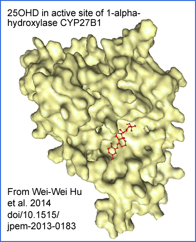
    The 25(OH)D molecule also relies on thermal vibrations and some local electrostatic attraction to rotate it into the correct orientation and so slot into the active site correctly.  I mention this to highlight that this reaction proceeds at a rate limited by the concentration of 25(OH)D, which is very low, and the likely still lower concentration of the 1-hydroxylase enzyme.  (Cofactors are necessary for the 1-hydroxylation reaction, some of which are consumed in the process.)

    The newly created 1,25(OH)2D molecule leaves the active site and the enzyme is ready for another 25(OH)D molecule to float into position.

    The rate at which 1,25(OH)2D production proceeds is directly dependent on the 25(OH)D level in the cytosol, and this is reduced for every 25(OH)D molecule hydroxylated to 1,25(OH)2D, or lost to 24-hydroxylase.

    For the intracrine signaling system to operate rapidly and fully, there needs to be a substantial rate of 25(OH)D to 1,25(OH)2D conversion, because the latter is short-lived.  This means there needs to be a continual supply of 25(OH)D to the cell, by diffusion from the interstitial fluid (or perhaps directly from the plasma).

    So the presence of adequate, externally supplied, 25(OH)D in the cytosol (which should be the case at all times - this presence not created by the CD46 initiation of this intracrine / paracrine signaling system) together with the newly created (by the initiation of the intracrine / paracrine signaling process) 1-hydroxylase enzyme leads directly to the outcome of this step: in-cytosol production of 1,25(OH)2D.

    Some of this 1,25(OH)2D is degraded by 24-hydroxylase - over time, most of it which remains in the cell will presumably be degraded in this way.  Some of this 1,25(OH)2D diffuses from the cell and may act on nearby cells as a paracrine agent.

    Some of this 1,25(OH)2D drives the next step.

  5. The newly created 1,25(OH)2D molecules in the cytosol soon find themselves bound to the VDR, for which they have a very high affinity.  There are a bunch of details regarding how the complex of the two "migrates"* to the nucleus, binds with other things and (by various mechanisms I am far from understanding - and I am not sure if anyone fully understands them all) the presence of large numbers of bound VDR complexes alters the transcription (AKA expression) of numerous genes.  The exact details of which genes are induced or suppressed, and to what degree, depends entirely on the cell type.

    * "Migration" is the usual term, but as far as I know there is no active, powered, directional mechanism for this.  Can anyone improve my knowledge?  I wrote "migrate" to be compatible with all I have read, but for now  I assume the bound complex diffuses around the place randomly, with some of them getting into the nucleus.

    This is the transcription phase of the intracrine signaling system. 

    There is some kind of degradation process for bound VDR complexes in the nucleus, so they don't accumulate and alter gene translation forever.  I don't know the details, but I guess the VDR itself is retained and is free to diffuse back into the cytosol.  However, there is surely a degradation process for the VDR as well, otherwise they would accumulate and this part of the intracrine signaling system would not be turned off when the initiating condition for the system is no longer active.  The same goes for the 1-hydroxylase enzyme, which I assume is degraded in some way at a modest rate.

  6. The changed mRNA numbers for the various genes, by altering the protein produced by ribosomes, alters the function of the cell.   This is the final output of this entire vitamin D driven intracrine signaling process for this particular type of cell. 

    mRNAs are rapidly degraded, and proteins (which may be exported or transformed in various ways) don't last forever.  So for the autocrine signaling system to continue to function in its activated state, the original initiating stimulus (in the Th1 cell type, C3b binding to the CD46 receptors bound to, and sticking out of, the cell's plasma membrane) must continue to be present, 25(OH)D needs to be continually supplied, by diffusion, so 1,25(OH)2D production can continue.  So there needs to be a continuing supply of 25(OH)D, by diffusion from the bloodstream into the cytosol of each cell.

    The exact details by which IFNγ and IL-10 are produced by these Th1 lymphocytes is somewhat more complex than getting their mRNAs into the cytosol.  More details of this were discovered by the researchers and are summarized below.  However, these details do not alter the central role of 25-hydroxyvitamin D based intracrine signaling controlling the Th1 cells' production of these two crucially important cytokines other than that they also rely to some extent on the hydroxyvitamin D based intracrine signaling system also upregulating the transcription of the gene which produces IL-6.

This has been a rather detailed excursion into the molecular details of the central (common to all cell types which use it) mechanisms of 25-hydroxyvitamin D based intracrine signaling. 

We need to think about this because at present (March 2021) the world is going to hell in a handbasket, in large part due to most humans having insufficient 25(OH)D in the cytoplasm of their immune cells to initially fight the SARS-CoV-2 infection, and in particular, in the context of humans' genetic predisposition to overly inflammatory immune responses due to lack of helminths:

https://aminotheory.com/cv19/#helminthsgone

(2023-06-29 update: on helminths, see: https://vitamindstopscovid.info/06-adv/ .

If everyone had about 50ng/ml or more - twice is just fine (2023-06-29 note: for most people, there may be some people who have bad reactions to 100 ng/mL 250 nmol/L, but I have not yet been able to find all the best research) - 25(OH)D in their bloodstream, all these immune cell types, Th1 lymphocytes included, and all other cells in the body which use 25-hydroxyvitamin D based intracrine (and perhaps paracrine) signaling, would be working pretty well.  Omega-3 and other nutrients such as magnesium, boron (#08-boron) and vitamin C are also important, but for now we focus on 25(OH)D, since this is the most important nutritional deficiency which clobbers all aspects of the immune system.

This McGregor et al. preprint concerns low 25(OH)D greatly increasing the (already genetically driven - see helminth material must mentioned) overly-inflammatory pattern of immune responses - and this response (perhaps just from these Th1 cells, but likely involving weak and dysregulated responses from all types of immune cell) is the primary driving reason why some COVID-19 sufferers progress to severe symptoms in their lungs, with high levels of endothelial damage, which causes the hypercoagulative blood which does the real damage with microembolisms and larger clots all through the body.

Inquiring minds now want to know how the (ideal, as far as we know ~50ng/ml 125nmol/L or more 25(OH)D blood levels relate to the levels of 25(OH)D in the cytoplasm of the Th1 cells (and all other cell types which also use vitamin D for autocrine / paracrine signaling).  See my pages at the current site and at https://vitamindstopscovid.info for research which shows we need such levels, at least for intracrine signaling to work properly. (Some people, with auto-immune diseases, need two or three times these levels to significantly reduce or abolish their symptoms - here I am discussing most people, not these.  See the McCullough et al. article mentioned above for examples.)

Looking at the experimenter's graphs for the IFNγ and IL-10 responses to 1,25(OH)2D (Fig 2C) and 25(OH)D (Fig S5d in the Supplementary data PDF), we see that most of the change occurs between concentrations of 0 and:

1,25(OH)2D: 1nM, with about half the action at 0.1nM.  (However, I chose 10nM for the levels which I think reflect the complete transition to anti-inflammatory state D.)

25(OH)D: (My guess, based on interpolating between the 0, 10 and 50nM levels.) 25nM with about half the action around 7nM.

nM == nano Mols == nmol/L = a billionth of a mole of molecules per litre.

1 nanogram per millilitre of D3, 25(OH)D or 1,25(OH)2D is the same concentration as 2.5nmol/L of these compounds.  (This is ignoring the slightly heavier mass of the latter two.)

A Mole [WP] is 6.022 x 10²³ molecules.  1 nanomol is 6 x 10¹⁴ molecules, so 1nM = 1nmol/L = 0.4ng/L means that for each molecule, there is 1.66 x 10⁻¹⁵ litres of water.  This volume is a cube with sides 1.185 micrometres = 1185 nanometres, which is big for a molecule which is only 0.2 nanometres long.   This is something like one 1mm long object in a home swimming pool.

The nM concentrations reported in these graphs are for the solution in which the researchers bathed the cells, and for now we assume reasonably free diffusion into the much smaller cell bodies - so these figures roughly represent the concentrations in the cytosol of these Th1 lymphocytes.

So it seems that to get the intracrine signaling system of these cells to respond fully, we need about 25nmol/L 25(OH)D in the cytosol, which is about 10ng/ml.

I think the discrepancy between this very approximate figure and the abovementioned ~50ng/mL for circulating 25(OH)D in the bloodstream can be accounted for roughly as follows:

  1. Most of the 25(OH)D in the blood plasma is bound tightly to vitamin D binding protein (DBP) molecules [WP], which with 458 amino acids, dwarf the 25(OH)D molecule which is not much bigger than a single amino acid.  There is is considerable individual and racial variation in the form of these DBP molecules and in the concentration in which they are present in the plasma.

    Although it is possible to measure the level of 25(OH)D which is not bound to DBP, this is not a commonly available from pathology labs, although such measurements might be more clinically relevant regarding the immune system.

    DBP also binds D3 and the low level of circulating (hormonal) 1,25(OH)2D.

  2. Most of the 25(OH)D which is not bound to DBP molecules is bound - much less strongly, I recall - to albumin proteins [WP].

    So the fraction of 25(OH)D available to diffuse into the interstitial fluid, and then into the cytosol of immune cells such as these Th1 lymphocytes, is the unbound fraction plus some part of the albumin-bound fraction.

  3. In any intracellular parts of the 25(OH)D molecules paths of diffusion from the plasma to the cytosol of the Th1 lymphocytes (such as diffusing through endothelial and other cells of capillaries), there will be some losses due to the actions of the 24-hydroxylase enzyme.
In March 2021 we know, from numerous research articles in the last year - many of which are analysed by some apparently expert, but anonymous, souls at: https://vdmeta.com , some of which are listed at https://aminotheory.com/cv19/#lr  - that people who succumb to severe COVID-19 have generally lower, on average, 25(OH)D levels than those who don't.   This becomes particularly clear when we consider the dramatic improvements due to giving hospitalised COVID-19 patients in Cordoba, Spain, just 0.532mg of oral 25(OH)D calcifediol: https://aminotheory.com/cv19/#2020-Castillo , raising their 25(OH)D levels from a likely 4 to 20ng/ml range to around 60ng/ml in a few hours.

So lets think about D3 intakes and these low 25(OH)D levels for a moment.  For 70kg adults, 0.125mg 5000IU D3 a day maintains about 50ng/ml.  This is 1 gram every 22 years.  These patients lower 25(OH)D levels of  4 to 20ng/ml probably result from much lower total quantities of D3 from UV-B, food and few, if any, supplements.  Let's say their D3 input was 0.01mg 400IU a day, with some patients, in recent years at least, with even less, due to being aged, being indoors, having dark skin, covering their bodies almost entirely when outside etc.  The UK government recommends 0.01mg 400IU D3 a day for adults - in winter only.

These people would generally not be patients in hospital with COVID-19 if they had been supplementing 0.125mg D3 a day (or more according to bodyweight, with more for people suffering from obesity: https://vitamindstopscovid.info/01-supp/ ).  2023-06:29: see the chart of vitamin D3 supplemental intake quantities, as ratios of body weight, at: https://brownstone.org/articles/vitamin-d-everything-you-need-to-know/ .

So this severe ill-health, and numerous other aspects of ill health, has been in large part caused by these patients having disastrously low D3 intakes (though generally about enough to ensure they don't get rickets or unusual levels of osteoporosis), of about 1 gram every 100,000 days = 274 years.  Pharma grade D3 costs about USD$2.50 a gram ex factory in 1kg quantities.  (That said, UK intracrine signaling pioneer Martin Hewison states in a 2021-03-03 video that the UK is the vitamin D deficiency centre of the world, and that rickets is not uncommon in certain communities - presumably of people with dark skin and/or who avoid direct sun exposure of their skin.)

This global epidemic of vitamin D deficiency has been running for decades and centuries.  (2023-06-23 update: since humans started living in Europe and especially northern Europe, eventually migrating to various other locations far from the equator.)  Without it, there would be little or no need for the disastrous lockdowns, social distancing, economic and travel shutdowns etc. which governments have imposed to combat COVID-19, and surely will continue to impose in the years as vaccine manufacturers (and populations of entire countries) play cat-and-mouse, or whack-a-mole, with  increasingly efficient viral variants which are also under strong selection pressure to evolve avoidance of existing infection- and vaccine-induced immune responses.

Back to the research article!

The researchers demonstrate further details of the Th1 immune shutdown program, which I discuss below.   This research shows that the shutdown is dependent on adequate 25(OH)D levels in the cytosol of the cell, since, in-vitro (as we surmise, in-vivo, from high 25(OH)D levels apparently causing a reduction in this hyper-inflammatory immune dysregulation) the cells do not respond to high levels of complement by transitioning from their pro-inflammatory program to their anti-inflammatory shutdown program, unless sufficient 25(OH)D is supplied to them.

This concludes the section of my summary and discussion which is most relevant to 25-hydroxyvitamin D based intracrine signaling in Th1 lymphocytes.  What follows is an attempt to summarize some of the other molecular details of exactly how IFNγ, IL-10 and IL-6 production is regulated in these cells, within the context of these Th1 cells' 25-hydroxyvitamin D based intracrine / paracrine signaling system.

Again, please remember this is the best effort of an electronic technician to understand and describe complex cell-biology experimentation.

The researchers noticed that IL-10 production was proportional to IL-6 production.  They added IL-6 to cells which had their CD46 receptors stimulated, and so the 25-hydroxyvitamin D based intracrine signaling system had been initiated, but with the cells remained stuck in their pro-inflammatory B state due to there being insufficient 25(OH)D in the cytosol.  (With sufficient 25(OH)D they would have converted some of it to 1,25(OH)2D, completed the autocrine signaling pathway and transitioned through C to the anti-inflammatory D state.)  This caused the cells to produce pro-inflammatory IL-17, which had been reported by other researchers.  This seems clinically relevant to me, since this is an additional mechanism for pro-inflammatory dysregulation in patients with low 25(OH)D levels, IL-6 being at high levels in all these COVID-19 lung infections. 

Both c-JUN and STAT3 transcription factors were produced by the full operation of the 25-hydroxyvitamin D based intracrine signaling system (leading to state D).  c-JUN phosphorylation [WP] was also driven directly by this.  STAT3 phosphorylation was dependent not on this  completion of the intracrine signaling pathway, but on the presence of IL-6, the production of which was  directly caused by the completion of the intracrine signaling system.  The researchers confirmed that the IL-6 receptor was necessary for this process by introducing an IL-6 receptor blocker Tocilizumab.  (Imagine the dreams of the people whose job it is to come up with these names for pharmaceuticals.)  

Since this IL-6 was, at least in part, produced inside the cell, and activates receptors in the same cell, this is a separate IL-6 based intracrine pathway with these IL-6 molecules acting as an intracrine agent.  To the extent that some of the IL-6 which activates these receptors was produced in nearby cells (Th1 or other types), those IL-6 molecules are acting as paracrine agents.

With further evidence from the cells of two patients with a rare STAT3 mutation (!) the researchers concluded:

VitD [they mean completion of the 25-hydroxyvitamin D based intracrine signaling response to high levels of complement, which is only possible with sufficient 25(OH)D] induces STAT3 and IL-6, and IL-6 phosphorylates STAT3 to promote production of [anti-inflammatory] IL-10.

Page 7 to 8 describes the researchers' investigation of the molecular details of how VDR activation (which will transition these cells from the B state, through C to D) works, such as by the VDR, complexed to other molecules, affecting the degree to which histones [WP] are acetylated [WP] in particular places.  The 46 chromosomes add up to 1.8 metres of DNA which is wound around much smaller histone molecules.  This typically tight winding impedes access to most of the DNA by the enzymes which can copy its information into messenger RNA molecules (mRNAs).  Acetylation in particular parts of the histone enables looser DNA winding and therefore greater access of these enzymes to particular parts of the DNA - including the genes which, in this particular cell type, VDR activation increases the transcription of.  I don't clearly understand their observations regarding c-Jun, BACH2 and STAT3 transcription factors, their phosphorylation and super enhancer structures arising from histone acetylation.

BACH2 - a transcription factor whose production is increased "by vitamin D" (meaning the successful completion of the 25-hydroxyvitamin D based intracrine process leading to state D) - is crucial to the increased transcription of other genes by this process, including the gene for the IL-6 receptor which, as as noted previously, is an essential link in the chain of IL-6 based intracrine / paracrine signaling which leads to the production of anti-inflammatory IL-10.

Expression of VitD-repressed genes . . . was significantly higher in bronchoalveolar fluid Th cells of  patients with COVID-19 than healthy controls.

So the Th1 cells from hospitalised COVID-19 patients did not have their (high levels of complement driven) CD46 activated 25-hydroxyvitamin D based intracrine signaling function run to completion (from state B through C to D), presumably due to lack of 25(OH)D, leading to little or no production of intracrine agent 1,25(OH)2D, and so little or no activation of VDRs which, if activated, would have repressed the transcription of these genes.

Here endeth the lesson. There will be a test on Friday!


#2020-Bilezikian

Review article on Vitamin D and COVID-19

Vitamin D and COVID-19
John P. Bilezikian, Daniel Bikle, Martin Hewison, Marise Lazaretti-Castro, Anna Maria Formenti, Aakriti Gupta, Mahesh V. Madhavan, Nandini Nair1, Varta Babalyan, Nicholas Hutchings, Nicola Napoli11, Domenico Accili, Neil Binkley, Donald W. Landry, Andrea Giustina  2020-08-04
https://eje.bioscientifica.com/view/journals/eje/aop/eje-20-0665/eje-20-0665.xml

This is a review of observations and mechanisms.  I haven't had a chance to read it yet.


#2020-Jalali-b

Farid Jalali's hypothesis of vascular damage and shunting via diffuse pulmonary-bronchial anastomoses

Be sure to read this beautiful detective work which offers the best explanation yet for many perplexing aspects of COVID-19.  The first link is to a video podcast with PDF and discussion and the second is to a text and graphic presentation with comments by Cameron Kyle-Sidell MD:

Scott Weingart. EMCrit Wee – A Theoretical Model of the Pathophysiology of COVID-19 with Farid Jalali (Not a Single Thing Verified–Pure Musings). EMCrit Blog. 2020-05-18

https://emcrit.org/emcrit/pathophysiology-of-covid19/  Excellent diagrams PDF

https://rebelem.com/covid-19-acute-lung-injury-a-proposed-model-v1-0-via-farid-jalali-md/


Cameron Kyle-Sidell's appreciative comments begin with:

I applaud Dr. Jalali for proposing a model for COVID-19 injury based on seemingly sound physiologic principles.  This model does provide explanations for anecdotal observations made by bedside physicians treating COVID-19 patients.  For example, the presence of dorsal-predominant shunting would explain why proning leads to marked oxygenation improvement that is not sustained once the patient returns to the supine position.

This work highlights the importance of protecting the integrity of the endothelium and limiting vasoconstriction, which is caused by excessive angiotensin II, resulting from SARS-CoV-2 destroying the ACE2 receptors.  See #2020-Kim below regarding how important vitamin D 1,25OHD is for protecting endothelial cells and inducing vasodilation

This is not necessarily just 1,25OHD in the plasma in general, but could be released in a paracrine fashion (short distance signaling between nearby cells [W]) after it is produced internally (in part for autocrine [W] signaling) and then diffuses out of the cell.  Autocrine cell signaling (Khan Academy explanation) is a big part of how immune cells control gene expression, and with leakage of the internally produced 1,24OHD, we have the possibility of paracrine reception of this on membrane-bound vitamin D receptors of nearby cells, such as the endothelial cells described by #2020-Kim , where it would upregulate endothelial protection and nitric oxide mediated vasodilation. 

What proportion of endothelial cell vitamin D receptors are on the apical surface, and so exposed to plasma 1,25OHD, plus any locally secreted by immune cells there?  What proportion are on the other side (outside the vessel or capillary = basolateral) and so, I guess less affected by the normal bone and calcium regulating level of 1,25OHD as a hormone, and more affected by the actions of immune cells in the surrounding tissue?

See also ../obesity/ According to the research I review there, the excess adipocytes of obesity express ACE2 receptors and so can be infected with SARS-CoV-2.  These are in the outer lining of the heart and there are also ectopic adipocytes in the cells which make up the alveoli.  I had never heard of these, but assuming it is true, then obese people would have further infection in their lungs combined with extra inflammation.

#2020-Jalali-c

Farid Jalali's hypothesis of serotonin (5-HT) playing a major role in COVID-19 lung pathology

In late June 2020, Farid Jalali released a text-only document:

http://www.jalali.one/farid/COVID19-Pathology.pdf

in which he proposes that serotonin, especially that released by platelets in the clotting cascade, plays a major role in making things much worse in SARS-CoV-2 infected lungs.  He suggests that the normal clearance mechanisms, which are in the lungs themselves, can no longer function, so serotonin builds up in the whole circulation causing further trouble.

As with the above diagrammatic hypothesis, I think only a handful of people - pulmonolgists and the like - could fully appreciate and critique what he is proposing.

However, I understand enough of it to envisage these as possible explanations for the extreme levels of damage which occur with the worst symptoms of COVID-19.

"Vitamin D" does not appear in either of these documents.  The big question is why is there so much inappropriate inflammation in these patients.  Surely low vitamin D levels - anything below 40ng/ml 250HD - plays a role in this immune system dysfunction.  I also suspect that low boron plays a role as well.  See: ../#08-boron and ../#boron-ra .


#2008-Hansdottir

Respiratory epithelial cells convert 25OHD to 1,25OHD

Further to the discussion above on intracrine and paracrine signaling of 1,25OHD, here is a pertinent article, from 2008.  The lead author is a critical care pulmonologist in Iowa.

Respiratory Epithelial Cells Convert Inactive Vitamin D to Its Active Form: Potential Effects on Host Defense
Sif Hansdottir et al. J Immunol November 15, 2008, 181 (10) 7090-7099
https://doi.org/10.4049/jimmunol.181.10.7090
Google Scholar 505 citations


. . . primary lung epithelial cells express high baseline levels of activating 1α-hydroxylase and low levels of inactivating 24-hydroxylase.  The result of this enzyme expression is that airway epithelial cells constitutively convert inactive 25-dihydroxyvitamin D3 to the active 1,25-dihydroxyvitamin D3.

Also, a 2011 article:

A review of the critical role of vitamin D in the functioning of the immune system and the clinical implications of vitamin D deficiency
Gerry K. Schwalfenberg Mol. Nutr. Food Res. 2011, 55, 96–108
https://doi.org/10.1002/mnfr.201000174 (Paywalled.)
https://sci-hub.tw/10.1002/mnfr.201000174
Google Scholar 265 citations

Toll like receptors (TLRs) respond to extracellular fragments of pathogens and/or of cellular damage, upregulate CYP27B1 (1a-hydroxylase) which converts 25OHD to 1,25OHD.  This activates the vitamin D receptor (the production of which is also upregulated by the activation of TLRs) and the activated receptor travels to the nucleus and upregulates genes specific to the particular cell type.  This diagram depicts some of the actions.  (I have another diagram somewhere depicting autocrine 1,25OHD signaling between cells.) 


He suggests 25OHD levels of 40 to 60ng, which I agree with.

#2020-Ackerman

Angiogenesis; ACE2-positive lymphocytes. alveolar and endothelial cells

Also of interest is this analysis of COVID-19 damage to lung tissue differs from that resulting from  H1N1 influenza in 2009:

Pulmonary Vascular Endothelialitis, Thrombosis, and Angiogenesis in Covid-19
Maximilian Ackermann et al. NEJM 2020-05-21
https://www.nejm.org/doi/full/10.1056/NEJMoa2015432

The lungs from patients with Covid-19 also showed distinctive vascular features, consisting of severe endothelial injury associated with the presence of intracellular virus and disrupted cell membranes. Histologic analysis of pulmonary vessels in patients with Covid-19 showed widespread thrombosis with microangiopathy. Alveolar capillary microthrombi were 9 times as prevalent in patients with Covid-19 as in patients with influenza (P<0.001). In lungs from patients with Covid-19, the amount of new vessel growth — predominantly through a mechanism of intussusceptive [splitting] angiogenesis — was 2.7 times as high as that in the lungs from patients with influenza (P<0.001).

Are these new blood vessels or capillaries fed by the pulmonary or bronchial artery? To what extent were they clogged with microthrombi?  To what extent could they participate in gas exchange?  Although the accompanying editorial notes that the H1N1 samples are not directly comparable, this graph indicates that angiogenesis is a very prominent feature in COVID-19.  This analysis is from autopsies - so this progression is for the patients with the worst symptoms.


79 inflammation-related genes were differentially regulated only in specimens from patients with Covid-19, whereas 2 genes were differentially regulated only in specimens from patients with influenza; a shared expression pattern was found for 7 genes.

This suggests to me a high immune system involvement, in addition to direct viral damage, in both the COVID-19 and H1N1 affected lungs.  Low vitamin D drives inappropriate, self-harming, overly-inflammatory responses, as well as weak antiviral defense mechanisms.

Loss of ACE2 receptors is a prominent part of Farid Jalali's hypothesis (above), driving excessive angiotensin II.  To what extent is this due to direct viral damage (virus attaches to ACE2 which is then internalised with the virus and so no longer remains active on the cell membrane) or due to immune system destruction of infected cells, and perhaps those nearby even if uninfected?

Why, in these illnesses are ACE2 receptors more frequently found on epithelial and endothelial cells?  Are these cells sensing excessive blood pressure due to microthrombi blocking the capillaries and/or vasoconstriction there?  If so, do they sprout ACE2 receptors in an attempt to reduce angiotensin II levels and so reduce vasoconstriction?

If so, then these extra ACE2 receptors make them vulnerable to viral infection and the whole situation can spiral out of control.

Fraction of cells with ACE2 receptors
Control
H1N1
CV19
Alveolar epithelial
0.05
0.35
0.25
Endothelial cells
0.07
0.55
0.49
Lymphocytes
0.00
0.15
0.22

Why do ACE2 receptors appear on lymphocytes?  Are these lymphocytes also acting to reduce local angiotensin II to reduce vasoconstriction?

If so, then the virus is well adapted: it gets into cells which already express the ACE2 receptor, taking these receptors, and then the cells, out of action.  This reduces angiotensin II levels, driving vasoconstriction and microembolisms which cause multiple cell types which normally express few, if any, ACE2 receptors to express lots of them, in an attempt to reduce vasoconstriction - so the virus can infect them too.  Inducing lymphocytes to express ACE2 and so be infected is quite a twist.

This article seems be relevant, mentioning ACE2-positive lymphocytes, but I have not attempted to pursue this line of inquiry further.  Please suggest pertinent articles and discussions.  ../#contact .

High expression of ACE2 receptor of 2019-nCoV on the epithelial cells of oral mucosa
Hao Xu et al. International Journal of Oral Science volume 12, Article number: 8 (2020)
https://www.nature.com/articles/s41368-020-0074-x
Google Scholar 249 citations


#2020-Kim

Vitamin D and Endothelial Function

Your COVID-19 patients are (according to Philippino and Indonesian research I cite here cv19/) generally seriously deficient in vitamin D.  Surely you can improve their chances of survival with vitamin D supplements. 

Cholecalciferol (D3) takes days to convert to 25OHD in the liver - and the liver may not be functioning well.  So high dose oral D3 - such as 50,000 or 100,000IU, ideally with a meal including fats - when they arrive in hospital, is one way to start.  If you can do oral of IV 25OHD (calcifediol = Rayaldee) then this would be faster.  Most immune system cells require plasma 25OHD for their internal synthesis of 1,25OHD to activate their internal (intracrine) vitamin D receptor signaling.  So the whole of immune regulation depends on a good 25OHD level. 

Other cells, such as endothelial cells, require 1,25OHD calcitriol at their membrane bound vitamin D receptors, and so may not rely directly on plasma 25OHD levels. 

Please see this recent review article:

Vitamin D and Endothelial Function
Do-Houn Kim et al. Nutrients 2020, 12(2), 575
https://www.mdpi.com/2072-6643/12/2/575

Adequate vitamin D activation of endothelial cells achieves numerous functions:
NO is a primary vasoactive substance that works as a potent vasodilator in addition to other vasoprotective properties such as protection from vessel inflammation and lesion formation.

Endothelium-derived NO acts on adjacent vascular smooth muscle cells in a paracrine manner and induces vascular muscle relaxation . . .

. . . protects the vessel from developing atherosclerosis by inhibiting platelet adherence and aggregation, and leukocyte activation.

#2020-Tay

The trinity of COVID-19: immunity, inflammation and intervention

Here is an excellent paper, from researchers in Singapore and Liverpool regarding the role of inappropriate, over-inflammatory, immune responses in severe symptoms with COVID-19.

There's no mention of vitamin D - though deficiency in this clearly drives weakened and dysregulated immune responses which allow disease progression and which, as this article documents, drive a lot of the damage which occurs.  Nor is there any mention of coagulation or vasoconstriction - both key elements in the material above, which was developed after this late April article would have been finalised.

The trinity of COVID-19: immunity, inflammation and intervention
Matthew Zirui Tay et al. Nature Reviews Immunology 2020-04-28
https://www.nature.com/articles/s41577-020-0311-8
Google Scholar 36 citations


. . . aggressive inflammatory responses strongly implicated in the resulting damage to the airways.  Therefore, disease severity in patients is due to not only the viral infection but also the host response.

In addition, the vast release of cytokines by the immune system in response to the viral infection and/or secondary infections can result in a cytokine storm and symptoms of sepsis that are the cause of death in 28% of fatal COVID-19 cases. In these cases, uncontrolled inflammation inflicts multi-organ damage leading to organ failure, especially of the cardiac, hepatic and renal systems.

Pyroptosis is a highly inflammatory form of programmed cell death that is commonly seen with cytopathic viruses.  A wave of local inflammation ensues, involving increased secretion of the pro- inflammatory cytokines and chemokines IL-6, IFNγ, MCP1 and IP-10 into the blood of afflicted patients.

Notably, there exists a highly inflammatory monocyte-derived FCN1+ macrophage population in the bronchoalveolar lavage fluid of patients with severe but not mild COVID-19.  Also, patients with severe disease show a significantly higher percentage of CD14+CD16+ inflammatory monocytes in peripheral blood than patients with mild disease. These cells secrete inflammatory cytokines that contribute to the cytokine storm, including MCP1, IP-10 and MIP1α.

Unrestrained inflammatory cell infiltration can itself mediate damage in the lung through excessive secretion of proteases and reactive oxygen species, in addition to the direct damage resulting from the virus.  Notably, virus was found in T lymphocytes, macrophages and monocyte-derived dendritic cells. Direct virus killing of lymphocytes could contribute to the observed lymphopenia in patients. 

Viral infection in immune cells such as monocytes and macrophages can result in aberrant cytokine production, even if viral infection is not productive. The degree to which SARS-CoV-2 targets these cells remains poorly defined.

Understanding the precise drivers of immune dysfunction is crucial to guide the application of appropriate immunomodulatory treatments.

Indeed.  The pages you are reading explain most or all of this immune system dysfunction in terms of individual genetic variation, lack of helminths, some dietary excesses and some very common nutritional deficiencies - especially vitamin D, boron, omega-3 fatty acids, vitamin C and potassium.

#2020-Teuwen

COVID-19: the vasculature unleashed

Here's another article concerning endothelial failure in the lungs, with the researchers not knowing why this happens to some people.

COVID-19: the vasculature unleashed
Laure-Anne Teuwen et al. Nat Rev Immunol (2020).
https://doi.org/10.1038/s41577-020-0343-0


I wonder who created this beautiful illustration.

During homeostasis, the endothelium, surrounded by mural cells (pericytes [W]), maintains vascular integrity and barrier function. It prevents inflammation by limiting EC - immune (EC = Endothelial Cell) cell and EC - platelet interactions and inhibits coagulation by expressing coagulation inhibitors and blood clot-lysing enzymes and producing a glycocalyx (a protective layer of glycoproteins and glycolipids) with anti-coagulation properties.

Interestingly, recent studies using single-cell transcriptomics revealed endothelial phenotypes that exhibit immunomodulatory transcriptomic signatures typical for leukocyte recruitment, cytokine production, antigen presentation and even scavenger activity. Compared with ECs from other organs, lung ECs are enriched in transcriptomic signatures indicating immunoregulation, and a subtype of lung capillary ECs expresses high levels of genes involved in MHC class II-mediated antigen processing, loading and presentation.

After the initial phase of viral infection, ~30% of hospitalized patients with COVID-19 develop severe disease with progressive lung damage, in part owing to an overreacting inflammatory response.

These would be mainly the patients with the weakest and most dysregulated immune systems - the primary, currently known (I suspect boron deficiency as well) preventable cause of which is vitamin D deficiency.  Yet vitamin D is not mentioned in this article.

Mechanistically, the pulmonary complications result from a vascular barrier breach, leading to tissue oedema (causing lungs to build up fluid), endotheliitis, activation of coagulation pathways with potential development of disseminated intravascular coagulation (DIC) and deregulated inflammatory cell infiltration.

Reduced ACE2 activity indirectly activates the kallikrein–bradykinin pathway, increasing vascular permeability.

Immune cells, inflammatory cytokines and vasoactive molecules lead to enhanced EC contractility and the loosening of inter-endothelial junctions.  In turn, this pulls ECs apart, leading to inter-endothelial gaps. Finally, the cytokines IL-1β and TNF activate glucuronidases that degrade the glycocalyx but also upregulate hyaluronic acid synthase, leading to increased deposition of hyaluronic acid in the extracellular matrix and promoting fluid retention.  Together, these mechanisms lead to increased vascular permeability and vascular leakage.

Vascular integrity and EC death leads to exposure of the thrombogenic basement membrane and results in the activation of the clotting cascade.

"Finally" just refers to this initial stage of the zombie apocalypse which overcomes all these cell types in the lungs.  Note that in the description so far, and in what follows, there is little or no mention of viral activity.  Most people deal with the virus pretty well, and things never get as bad as just described.  If you have read this page and most of the ../cv19/ page you will be at least roughly aware of the protective, largely vitamin D dependent mechanisms in these cells which protect them from damage by pathogens, and disallow the storm of destruction described so well here, which is primarily driven by a weakened and dysregulated immune response.

Moreover, ECs activated by IL-1β and TNF initiate coagulation by expressing P-selectin, von Willebrand factor and fibrinogen, to which platelets bind. In turn, ECs release trophic cytokines that further augment platelet production. Platelets also release VEGF, which triggers ECs to upregulate the expression of tissue factor, the prime activator of the coagulation cascade, which is also expressed by activated pericytes.  In response, the body mounts countermeasures to dissolve fibrin-rich blood clots, explaining why high levels of fibrin breakdown products (D-dimers) are predictive of poor patient outcome. As a result of the DIC and clogging/congestion of the small capillaries by inflammatory cells, as well as possible thrombosis in larger vessels, lung tissue ischaemia develops, which triggers angiogenesis and potential EC hyperplasia.  While the latter can aggravate ischaemia, angiogenesis can be a rescue mechanism to minimize ischaemia.  However, the newly formed vessels can also promote inflammation by acting as conduits for inflammatory cells that are attracted by activated ECs.

We're not done yet . . .

Many patients with severe COVID-19 show signs of a cytokine storm. The high levels of cytokines amplify the destructive process by leading to further EC dysfunction, DIC, inflammation and vasodilation of the pulmonary capillary bed.    

This explanation differs from that of Farid Jalali's #2020-Jalali-b above - in which angiotensin II levels force vasoconstriction.  The above words seem to indicate that the unspecified mechanisms of vasodilation (I guess nitric oxide, as noted above) prevail. 

This results in alveolar dysfunction, ARDS with hypoxic respiratory failure and ultimately multi-organ failure and death

EC dysfunction and activation likely co-determine this uncontrolled immune response


Yes, but why?  This is where anyone who has read the research I have read - and which you will read if you follow my links - thinks of the low 25OHD levels of these patients, and how this will reduce the ability for numerous types of immune cell (and it seems we should count some or all endothelial cells as immune cells too) to act strongly and successfully against the virus, while regulating the destructive responses which cause almost all the harm described in this article.
 
This is because ECs promote inflammation by expressing leukocyte adhesion molecules, thereby facilitating the accumulation and extravasation of leukocytes, including neutrophils, which enhance tissue damage.  This is because ECs promote inflammation by expressing leukocyte adhesion molecules, thereby facilitating the accumulation and extravasation of leukocytes, including neutrophils, which enhance tissue damage [Neutrophils are one of the immune system's professional phagocytes. W  Moreover, we hypothesize that denudation of the pulmonary vasculature could lead to activation of the complement system, [W] promoting the accumulation of neutrophils and pro-inflammatory monocytes that enhance the cytokine storm

As I wrote on the main page cv19/ most of the death resulting from influenza is also due to weakened, dysregulated, immune systems, with the same primary causes (cv19/#helminthsgone): lack of helminths, individual genetic variation, dietary excesses and nutritional deficiencies, most particularly of vitamin D.
This is based on the observation that during influenza virus infection, pulmonary ECs induce an amplification loop, involving interferon-producing cells and virus-infected pulmonary epithelial cells. Moreover, ECs seem to be gatekeepers of this [destructive and quite likely deadly] immune response, as inhibition of the sphingosine 1 phosphate receptor 1 (S1PR1) in pulmonary ECs dampens the cytokine storm in influenza infection. This raises the question whether pulmonary ECs have a similar function in the COVID-19 cytokine storm and whether S1PR1 could represent a therapeutic target. Another unexplained observation is the excessive lymphopenia in severely ill patients with COVID-19 and whether this relates to the recruitment of lymphocytes away from the blood by activated lung ECs.

Yes they are a therapeutic target - along with every other type of cell in our bodies.  Everyone should run their bodies better by giving all their cells the nutrients they require for proper operation.  This starts with 40 to 60ng/ml 25OHD.

When they don't do this, and their levels are 30ng/ml or less, sometimes down to 10ng/ml or less (and see ../#21authors for an article written by 21 experts who regard vitamin D deficiency as being only below 10ng/ml) they can get by - until a virus, bacteria or some other insult triggers this weak and dysregulated immune response.  Then they suffer sepsis, pneumonia etc. - or in the case of COVID-19 and even worse horror story because of the way the virus, during this self-destructive cascade, infects immune cells which respond to the troubles by expressing ACE2 receptors, driving a hypercoagulative storm which wreaks likely permanent harm all over the body.

When people get to hospital, in these conditions, the first thing doctors should do is try to raise their 25OHD levels to something healthy, like 40ng/ml.  Perhaps it will be too late, but I am exceedingly perplexed, spending these months knowing how low 25OHD is not being treated, and is condemning hundreds of thousands of people to terrible suffering, lasting harm and death.

We should remember that the wholesale 1kg ex-factory cost of D3 for 4000IU a day is US 9 cents per year.

All these people are boron deficient too.  Boron's half life is 20 hours or so, and they start of boron deficient in ordinary life, getting about 1mg or so a day, and become even more deficient in hospitals since doctors don't even recognise it as a nutrient.  Yet boron has numerous anti-inflammatory properties, similar to vitamin D, but also in some ways unique to itself.

100mg of borax a day gives 11.4mg boron a day, which is surely sufficient for repletion.  This is 36 grams of borax a year, which costs (retail, USD$10/kg) US 36 cents per year.

Additional circumstantial evidence suggests a link between ECs, pericytes and COVID-19. First, risk factors for COVID-19 (old age, obesity, hypertension and diabetes mellitus) are all characterized by pre-existing vascular dysfunction with altered EC metabolism.  As hijacking of the host metabolism is essential for virus replication and propagation, an outstanding question is whether EC subtypes or other vascular cells in specific pathological conditions have a metabolic phenotype that is more attractive to SARS-CoV-2.

Old age - in the absence of 4000IU or more D3 a day supplementations - is strongly correlated with even lower 25OHD levels than is normal middle age.  These risk factors are caused, in large part, by vitamin D and other deficiencies, the lack of helminths etc. and excessive dietary fats, sugars and other carbohydrates, and frequently lack of proper exercise. 

So even if the actual conditions were not risk factors in themselves (and they are, especially obesity) then the correlation between these conditions and risk for serious COVID-19 symptoms would be to a large extent explained by a lifetime of inadequate vitamin D.

Second, occasional clinical reports suggest an increased incidence of Kawasaki disease, a vasculitis, in young children with COVID-19.

I had never heard of Kawasaki disease.  It took me less than a minute with Google Scholar, searching for it with "vitamin D" to find the article I link to here ../#2015-Stagi which shows that the only children who suffer and die from this terrible condition have exceedingly low (rickets levels) of 25OHD.

Other articles on COVID-19 endothelial pathology

There are no doubt other articles with detailed accounts of COVID-19 pathology in the epithelium of the lungs.  I don't have time to find or read them all, but here are some of potential interest:

Overcoming Barriers The Endothelium As a Linchpin of Coronavirus Disease 2019 Pathogenesis
Dakota Gustafson et al. Arterioscler Thromb Vasc Biol. 2020;40:00–00

https://www.ahajournals.org/doi/pdf/10.1161/ATVBAHA.120.314558

Coronavirus Disease 2019 Coagulopathy: Disseminated Intravascular Coagulation and Thrombotic Microangiopathy - Either, Neither, or Both
Marcel Levi and Jecko Thachil Semin Thromb Hemost 2020-06-08

https://www.thieme-connect.com/products/ejournals/pdf/10.1055/s-0040-1712156.pdf


#vit-K

Vitamin K depletion in severe COVID-19 contributing to coagulation?

I haven't read this yet.  Would vitamin K supplementation reduce the most destructive aspect of severe COVID-19: the hypercoagulative state?

Reduced Vitamin K Status as a Potentially Modifiable Prognostic Risk Factor in Covid-19
Anton S.M. Dofferhoff et al.

https://www.preprints.org/manuscript/202004.0457/v2


#2020-Front #2020-FLCCC

MATH+ Protocol for early intervention to reduce cytokine storm and so reduce or eliminate the deadly hypercoagulative state

The website of the Front Line COVID-19 Critical Care Alliance is:

https://flccc.net

(Previously Front Line COVID-19 Critical Care Working Group  covid19criticalcare.com and before that: recoverywithoutwalls.com/covidprotocol/ .)

Please follow all the links to protocol, press release and other documents there, as well as documents linked from theirs:

General Adaptation in Critical Illness: Glucocorticoid Receptor-alpha Master Regulator of Homeostatic Corrections
Meduri & Chrousos
https://www.frontiersin.org/articles/10.3389/fendo.2020.00161/full

Outcomes of Metabolic Resuscitation Using Ascorbic Acid, Thiamine, and Glucocorticoids in the Early Treatment of Sepsis
Jose Iglesias et al.
https://www.sciencedirect.com/science/article/abs/pii/S0012369220304554

https://sci-hub.tw/10.1016/j.chest.2020.02.049


A 2018 article about this by Paul Marik and colleagues: Ascorbic acid, corticosteroids, and thiamine in sepsis: a review of the biologic rationale and the present state of clinical evaluation  www.ncbi.nlm.nih.gov/pmc/articles/PMC6206928/ contains a big diagram and explanation.

The protocol concerns the early use of methylprednisolone, intravenous vitamin C, thiamine (), low molecular weight heparin and other compounds, including vitamin D and melatonin to reduce or prevent the development of the cytokine storm which causes the hypercoagulative state which is what harms and kills people.  Other elements are nasal cannula oxygen with prone positioning and avoidance of incubation as much as possible.  


#Marik-protocol

The Marik Protocol

Paul Marik MD is a member of the FLCCC Alliance mentioned above.  His work predates COVID-19 and is applicable to most ICU patients, especially those with sepsis and lung injury.  His protocol is available from:

https://www.evms.edu/covid-19/medical_information_resources/

Paul Marik's approach was discussed in March 2017 at  https://emcrit.org/pulmcrit/metabolic-sepsis-resuscitation/ .  At that page, the second video is of some nurses at the East Virginia Medical School hospital.  At 1:09, Team Coordinator Kathi Hudkins recalls her team's initial thoughts on Dr Marik's protocols:

"This had to be some soft of fallacy.  It just seemed too simple."

But then their patients started getting better.

Despite the obvious necessity of supplementing a micronutrient which is essential to immune function, Paul Marik faces an uphill battle having his ideas accepted - and he considers it unethical to do an RCT, since half his patients would be denied a treatment which he knows works.

Dr Paul Marik has written over 400 peer-reviewed journal articles, 50 book chapters and 4 critical care books.  In 2017 criticalcarereviews.com/...cure-for-sepsis-with-paul-marik he said:

My colleagues, when I told them what was going on, thought it was  the biggest load of nonsense they had ever seen.  But then they actually saw that none of our patients were dying.  And then our CEO saw that none of the patients were dying.  So this has become instituted through the whole healthcare system.

"We were going to do a randomised clinical trial, but we couldn't - because it would have been unethical."

At this page there is also a 2020-05-28 video - which is actually at YouTube - in which Professor Marik discusses COVID-19 in general and how he and his team treat it.  His part in the video starts at 45 minutes. 

At 50:30 he mentions vitamin D status as one of the factors affecting COVID-19 outcomes - and the effect of high geographical latitude on low vitamin D levels.  In US states north of 40° there is a much higher risk of COVID-19 mortality.

COVID-19 results in a very high expression of inflammatory cytokines, chemokines, IL-6 and IL-1  [much more so than common respiratory viruses, including influenza]. 

What makes this virus so unique and so smart is that while it upregulates the inflammatory response, it downregulates the expression of interferons. 
Interferon type 1 and type 3 are the major host defenses against viral infections.  COVID-19, as opposed to influenza, results in a limited antiviral response. So there is a marked imbalance between the host immune response in getting rid of the virus and this profound inflammatory response.


EMCrit discussion on timing of corticosteroids

Josh Farkas and commenters discuss the timing and strength of corticosteroid use with COVID-19:

https://emcrit.org/pulmcrit/steroid-covid/

Dr Cameron Kyle-Sidell's videos on how different COVID-19 is from what he trained for

His impassioned video:  https://www.youtube.com/watch?v=g3ka8lo_fZ8 .  A later interview with John Whyte in which he mentions that his view of what was good for the patient was incompatible with the guidelines followed by the rest of his ICU team, so he resigned his position to work in the ER: https://www.medscape.com/viewarticle/928156 .

L. Gattinoni et al. on L and H "phenotypes"

https://www.esicm.org/wp-content/uploads/2020/04/684_author-proof.pdf


#2020-Jalali-a

Dr Farid Jalali's COVID-19 diagram concerning cytokine storm and resulting hypercoagulative state


Farid Jalali https://twitter.com/farid__jalali tweeted a detailed diagram https://twitter.com/farid__jalali/status/1247036001349849088 depicting the cytokine storm turning a moderately PAI-1 elevated coagulative state, which apparently can be controlled with heparin, into a hypercoagulable Progressive Thrombotic Cascade.  The long progression of this thrombosis apparently enables the oxygen levels to drop slowly enough that the patient copes with it better than would be the case with normal ARDS.  This leads to microvascular thrombosis in the lungs, heart, kidney, gut, pancreas, skin and CNS - and so frequently to death.

He states that COVID-19 is cytopathic to the endothelium and that this degenerates (as far as I know, in the absence of vitamin C etc. treatment) into the Severe COVID-19 cytokine storm.  This makes me think that the vitamin C, corticosteroid etc. treatment prevents the virus and the consequent (at least in these patients) immune response doing so much harm to the endothelium.


../ to the main COVID-19 page of this site.

To the index page of this site: ../../
Contact details and copyright information: ../../contact/
© 2020 Robin Whittle - please link to this site rather than copy the whole of its contents    Daylesford, Victoria, Australia